Advertisement
Advertisement
September/October 2026
Sponsored by Penumbra, Inc.
Pulmonary Embolism in 2026: New Guidelines, Data, and Treatment Pathways
With Timothy Fernandes, MD; Suhail Dohad, MD; Sanah Christopher, MD; Jason Mader, DO; and Michael Bode, MD
Pulmonary embolism (PE) care is entering a new chapter. In February 2026, a multisociety guideline introduced an A to E clinical classification framework and gave class I support for PE response team (PERT) assessment to improve in-hospital care—the first major update since the 2019 European Society of Cardiology guidelines.1,2
Because the guideline review preceded the first randomized mechanical thrombectomy trials, emerging evidence must be interpreted alongside its recommendations. In STORM-PE, category C3 patients randomized to anticoagulation plus Computer Assisted Vacuum Thrombectomy (CAVT®) with Lightning Flash® (Penumbra, Inc.) had a greater 48-hour right ventricular/left ventricular (RV/LV) ratio reduction than anticoagulation alone (29.7% vs 13.1%; P < .001), with 4.3% versus 7.5% composite major adverse events, no device-related transfusions, and 100% technical success.3* At 90 days, CAVT patients also had better functional outcomes, walking 479 versus 368 m on the 6-minute walk test (P = 0.049) and achieving New York Heart Association class I status in 97% versus 76% of patients (P = 0.02).4
*STORM-PE was only powered for primary endpoint of change in RV/LV ratio.


In this Q&A, Drs. Timothy Fernandes and Suhail Dohad discuss how they are applying new guidance and emerging evidence in practice—from PERT design and patient stratification to mechanical thrombectomy, discharge planning, and long-term recovery.
What does your PERT consist of, and what is your perspective on PERTs?
Dr. Dohad: PERTs have now become a class I recommendation in the new guidelines. That recommendation was included because of growing evidence that PERTs can improve the care and throughput of patients with PE. Part of the benefit is earlier patient identification and a more systematic approach to evaluation and management. In my experience, there is definitely a higher use of catheter-directed therapies among patients evaluated by PERTs given the evidence demonstrating shorter lengths of stay, improved outcomes, and, in some circumstances, possible mortality benefit.
PERTs are essential not only for acute care but also for discharge planning and longitudinal follow-up. Care becomes much more organized and protocol driven. Patients are followed more carefully, which can improve medication adherence, help them understand their disease and what to expect, and give them a reliable resource for PE- and medication-related questions.
Dr. Fernandes: Every hospital has different resources and interested parties. At University of California San Diego (UCSD), our PERT is primarily the pulmonary critical care doctors, who are the first call. We have a platform from Aidoc that alerts us to a PE, so we see cases come across and know the patient is there in the emergency room. It facilitates communication with whoever is on call for our interventional radiologists, and we can decide which intermediate- and high-risk (class C-E) patients need an intervention and treat them quickly. We have the capability of using extracorporeal membrane oxygenation for our sickest patients, so our cardiothoracic surgeons are involved with those decisions, and we can loop in our intensivists. How you design your PERT will depend on what resources you have available.
How do noninterventional members participate, and how is information communicated?
Dr. Dohad: Most of our PERT members are noninterventionalists. The broader team includes hematology, neurology, cardiac surgery, and thoracic or cardiothoracic surgery. That said, we keep the initial conversation streamlined. When a PERT call comes in, we do not bring in every specialist unless there is a specific clinical need. The initial discussion usually involves the pulmonary critical care attending, the pulmonary fellow, my fellow (a senior complex coronary, interventional endovascular fellow) and me.
Typically, a patient is presented through a group message. We review the initial presentation and all available imaging, order any imaging that is missing, and assess the patient’s hemodynamics: heart rate, blood pressure, respiratory rate, and overall clinical status. We also review key laboratory data, including biomarkers, lactate, hemoglobin, and other pertinent results. We discuss whether the PE appears provoked or unprovoked, as well as comorbidities that affect bleeding risk, cancer management, prognosis, and the patient’s underlying disease burden.
From there, we decide whether intervention is needed immediately or can be delayed until the next day, or whether the patient should be followed medically—and we then determine the most appropriate therapy, whether that is mechanical thrombectomy, catheter-directed thrombolysis, anticoagulation alone, or watchful waiting.
Dr. Fernandes: One of the things that makes the UCSD model a little different is that the pulmonary vascular specialist goes to the lab with our interventional radiologist. Decisions about when to stop or whether to go a little bit more are mutual decisions. That echoes the whole PERT model: This is multidisciplinary and collaborative, and deciding who to treat and when you’re finished can also be multidisciplinary.
How do you risk stratify, and what tools do you rely on?
Dr. Fernandes: There are a lot of tools available—Hestia criteria, Simplified PE Severity Index (sPESI), PE Severity Index (PESI), Bova score, etc. I think the composite PE severity (CPES) score is best as it includes the six things I care about: the two biomarkers, troponin and pro-B-type natriuretic peptide (proBNP); central PE; RV/LV ratio > 1 or RV dilatation; concomitant deep vein thrombosis (DVT)—often preluding an imminent second event and risking deterioration; and tachycardia. If your heart rate is > 100 bpm, you’re using the one trick you have: Cardiac output equals stroke volume times heart rate. If your heart rate is fast, the next thing to go is your cardiac output, and you start syncopizing and arresting. In the new guidelines, we really focus on categories C, D, and E.
Dr. Dohad: We use risk-assessment tools, including CPES score and NEWS2 (National Early Warning Score 2) score, to determine where the patient falls within the framework of the new guidelines—for example, C2, C3 with risk, D1, or D2 with risk. We consider central clot burden and hemodynamics when determining whether thrombectomy or another catheter-directed procedure is indicated and how urgently it should be performed.
Which patients do you select for mechanical thrombectomy, and what data inform that decision?
Dr. Dohad: Mechanical thrombectomy is generally our primary therapy because we believe it can provide the most effective result in the shortest time. The safety of the procedure has been demonstrated not only in randomized trials, including STORM-PE,3 but also in STRIKE-PE (NCT04798261),5 a large prospective registry of > 1,000 patients. STRIKE-PE includes core-lab adjudication and systematic follow-up, including quality-of-life measures, patient-reported outcomes, and 6-minute walk test. These data give us a clearer sense of what to expect after thrombectomy, including likely response and 30-day, 90-day, and 1-year outcomes. It makes the decision-making process more evidence-based.
Dr. Fernandes: Symptom onset > 14 days is an exclusion criterion for a lot of these interventional trials. We feel like we can identify chronic clot when we see it, but in the German FOCUS-PE trial, 15% of patients enrolled in this acute PE long-term follow-up study actually had chronic clot at presentation.6 It’s much more prevalent than we think.
When you’re looking at a CT for a patient who you’re considering taking to the lab, always stop and ask: Is this the acute presentation of chronic thromboembolic disease or chronic thromboembolic pulmonary hypertension, or is this acute PE? Acute PE you can help. If you go after chronic clot that has scarred into the vessel wall, you can get into a lot of trouble. Take the pressures into account as well. A right heart is not able to generate very high pressures acutely. If you’re seeing pulmonary artery (PA) systolic pressures in the 70s, 80s, or 90s, that’s a heart that has been seeing that pressure for a long time and has remodeled. So, if the pressure is very high, really hit pause and think about what you’re doing.
What does recovery look like, and how are you thinking about discharge and long-term outcomes?
Dr. Fernandes: I commend Drs. Lookstein and Rosovsky for the STORM-PE trial because it was the first big trial to look at a patient-centered outcome beyond did you live, did you die, or did you need the intensive care unit (ICU). We had a recent publication looking at the long-term sequelae of PE, and 12% of patients had evidence of chronic clot and an exercise limitation identifiable on cardiopulmonary exercise testing, along with chronic symptoms.7 One in eight patients after a PE will have these persistent exercise limitations. If we’re only thinking about who is going to live or die, then when a patient comes back to see me in clinic with a clot that could have been intervened upon and is now symptomatic with chronic clot, I’m kicking myself that we didn’t do something sooner. With the STORM-PE data, and the soon-to-follow PE-TRACT data (NCT05591118), the question becomes: Does being more aggressive with the acute presentation of PE prevent these long-term sequelae? I think it absolutely must.
What comes next in the PE space?
Dr. Dohad: The first priority is helping every hospital that treats PE become more organized. As more data emerge, we will need to digest and collate those data sets, determine which findings follow consistent trends, and understand any discordant results. We will also gain more information about long-term outcomes. The most important priority is making PE care accessible, not only within the United States but around the world. We have made a major impact over the last 10 years, but the work has just begun. A large proportion of people globally still do not have access to organized PE care. As programs become more organized and therapies become more available, training will be essential. It has taken us 10 years to become proficient, so there will likely be a learning curve as PE programs expand.
Dr. Fernandes: This is an exciting time in the PE space, with a lot of innovation and a number of ongoing clinical trials. We’ve really moved the needle on treating the sickest patients, but the next round of clinical trials will increasingly focus on patient-centered outcomes. We saw this in the longer-term STORM-PE data, where patients treated with catheter-directed thrombectomy had an approximately 100-m improvement in 6-minute walk distance.4 That is really important to patients because persistent shortness of breath is something they often report when returning to clinic after PE. We’re not only moving the needle on PE-related mortality; we can also help patients with long-term recovery after PE.
Disclosures:
Dr. Fernandes: Consultant to Penumbra®.
Dr. Dohad: Consultant to Penumbra®.
1. Writing Committee Members, Creager MA, Barnes GD, Giri J, et al. 2026 AHA/ACC/ACCP/ACEP/CHEST/SCAI/SHM/SIR/SVM/SVN guideline for the evaluation and management of acute pulmonary embolism in adults: a report of the American College of Cardiology/American Heart Association Joint Committee on Clinical Practice Guidelines. Circulation. 2026;153:e977-e1051. doi: 10.1161/CIR.0000000000001415
2. Konstantinides SV, Meyer G. The 2019 ESC guidelines on the diagnosis and management of acute pulmonary embolism. Eur Heart J. 2019;40:3453-3455. doi: 10.1093/eurheartj/ehz726
3. Lookstein RA, Konstantinides SV, Weinberg I, et al. Randomized controlled trial of mechanical thrombectomy with anticoagulation versus anticoagulation alone for acute intermediate-high risk pulmonary embolism: primary outcomes from the STORM-PE trial. Circulation. 2026;153:21-34. doi: 10.1161/CIRCULATIONAHA.125.077232
4. Lookstein R. Clinical, functional, and quality of life outcomes through 90 days in the STORM-PE RCT for mechanical thrombectomy with anticoagulation vs anticoagulation alone in acute intermediate-high risk PE patients. Presented at: Society of Interventional Radiology Annual Scientific Meeting (SIR); April 11-15, 2026; Toronto, Canada.
5. Konstantinides S, Moriarty JM, Dohad SY, et al. STRIKE-PE interim analysis: one-year functional outcomes and quality of life after treatment of pulmonary embolism with computer assisted vacuum thrombectomy. Eur Heart J. 2026;15 (Supp):i428-1430. doi: 10.1093/ehjacc/zuag046.246
6. Valerio L, Mavromanoli AC, Barco S, et al. Chronic thromboembolic pulmonary hypertension and impairment after pulmonary embolism: the FOCUS study. Eur Heart J. 2022;43:3387-3398. doi: 10.1093/eurheartj/ehac206
7. Morris TA, Dakaeva K, Alotaibi M, et al. Chronic thromboembolism phenotypes after acute pulmonary embolism: identification with the SEARCH algorithm. Eur Respir J. 2026;68:2501562. doi: 10.1183/13993003.01562-2025
Acute PE With RV Dysfunction Treated With Lightning Flash 3.0

PATIENT PRESENTATION
A man in his late 50s with gastroesophageal reflux disease presented with syncope and acute dyspnea after a long-haul flight. He was normotensive but tachycardic (105 bpm) and required 4 to 6 L/min nasal oxygen to maintain a peripheral capillary oxygen saturation of 90% to 95%. An electrocardiogram (ECG) showed sinus tachycardia.
Troponin I was elevated at 191 ng/L and proBNP at 134 pg/mL. Echocardiography showed RV enlargement and dysfunction (tricuspid annular plane systolic excursion, 1.4 cm; RV/LV ratio, 2.5). PESI was 108 and CPES was 6. Given symptomatic acute PE with hypoxemia, biomarker elevation, and RV dysfunction, the multidisciplinary team classified him as American Heart Association (AHA)/American College of Cardiology (ACC) D1/D2 and proceeded with catheter-based therapy.
INTERVENTION AND OUTCOME
CAVT was performed with Lightning Flash 3.0 (Penumbra, Inc.). The patient’s baseline PAP was 32/12 mm Hg (mean, 20 mm Hg), with a heart rate of 100 to 120 bpm. The 35-minute procedure included 8 minutes of device time (Figures 1, 2 and 3). After thrombectomy, the patient’s PAP decreased to 23/10 mm Hg (mean, 15 mm Hg), his heart rate fell to 60 to 70 bpm, and he maintained 100% oxygen saturation at 2 L/min oxygen.
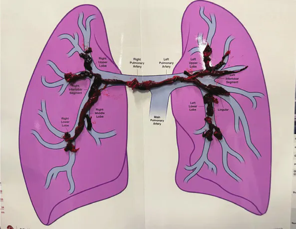
Figure 1. Thrombus aspirated.
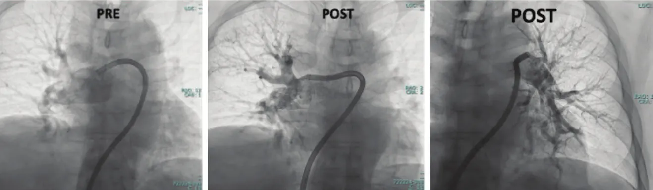
Figure 2. Pre- and post-thrombectomy pulmonary angiography.
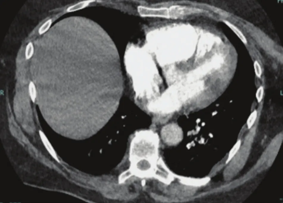
Figure 3. Preprocedural CT pulmonary angiography.
TAKEAWAY
This case demonstrates immediate hemodynamic and clinical improvement after thrombectomy and underscores the value of integrating symptoms, oxygen needs, biomarkers, RV function, and hemodynamics when considering escalation of care.
Acute Bilateral PE With Right Heart Strain Treated With Lightning Flash 3.0

PATIENT PRESENTATION
A woman in her mid 30s with obesity, oral contraceptive use, and a family history of venous thromboembolism presented with tachycardia, dyspnea, near syncope, hypotension, and hypoxemia. CTA showed extensive bilateral PE with right heart strain (RV/LV ratio of 1.5, RV dysfunction, positive McConnell sign) (Figure 3). Troponin was elevated to 1,186 ng/mL, consistent with AHA/ACC stage C3R acute PE.
INTERVENTION AND OUTCOME
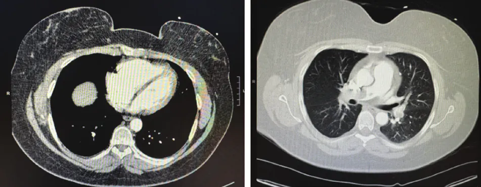
Figure 3. Preprocedural CT pulmonary angiography.
Mechanical thrombectomy was performed with the Penumbra Indigo® Lightning Flash 3.0 system. An occlusive right iliac vein thrombus identified on femoral access prompted conversion to right internal jugular access and placement of an inferior vena cava filter. The Element™ sheath was utilized for access, traversed into the main PA, followed by the introduction of Lightning Flash 3.0. Using the Element Assist technique (Penumbra, Inc.), acute thrombus was removed from the right PA and lobar branches and the left lower lobe PA (Figure 1).
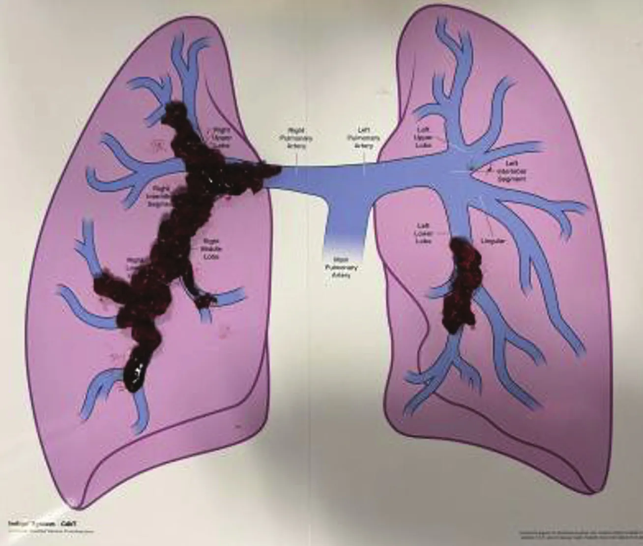
Figure 1. Thrombus aspirated.
Angiography showed near-complete thrombus removal and restored perfusion (Figures 2 and 3). The mean PAP decreased from 31 to 15 mm Hg, heart rate decreased from 126 to 96 bpm, blood pressure stabilized, and oxygen saturation improved from 90% on 4 L/min oxygen to 98%. The total procedure time was 66 minutes from access to closure, with a 50-mL estimated blood loss and no reported complications.
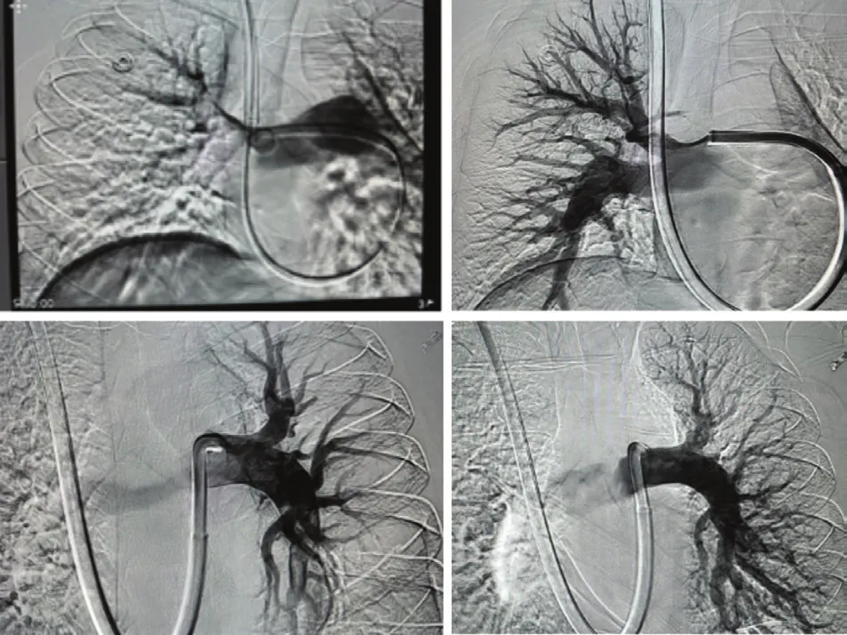
Figure 2. Pre- and post-thrombectomy pulmonary angiography.
TAKEAWAY
The patient did not require ICU admission, was weaned from oxygen, and was discharged to home after 2 days on apixaban, with outpatient management planned for a right iliac DVT.
Lightning Flash 3.0 Thrombectomy for Intermediate-High–Risk PE in a Patient With Multiple Comorbidities

PATIENT PRESENTATION
A man in his early 70s with hypertension, hyperlipidemia, diabetes mellitus, and neuropathic pain presented with a 2-day history of dyspnea. On admission, he was tachycardic (heart rate, 105 bpm) and hypoxemic (oxygen saturation, 86%). Laboratory testing demonstrated elevated troponin (250 ng/L), BNP (404 pg/mL), and D-dimer (13,197 ng/mL). Lower extremity ultrasound identified a partially occlusive thrombus in the left posterior tibialis vein.
CTA demonstrated bilateral main, lobar, and segmental PE with a large thrombus burden and evidence of right heart strain. The RV/LV ratio was borderline elevated at 0.93. With an sPESI score of 2, elevated cardiac biomarkers, hypoxemia, and imaging evidence of RV strain, the patient was classified as having intermediate-high–risk PE consistent with AHA class C2/3. Prior to intervention, the patient remained normotensive with a heart rate of 90 bpm and an oxygen saturation of 94% while receiving 7 L high-flow nasal cannula oxygen. The invasive PAP was 54/19 mm Hg (mean, 34 mm Hg). A preprocedural echocardiogram was not available because of the urgent need for intervention.
INTERVENTION AND OUTCOME
The patient underwent Computer Assisted Vacuum Thrombectomy (CAVT) via right femoral venous access using a 17-F, 65-cm Element Sheath (Penumbra, Inc.) (Figure 1).
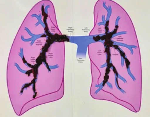
Figure 1. Thrombus aspirated.
Thrombectomy was performed with the Lightning Flash 3.0 system with CAT™16 (Penumbra, Inc.). The total case time was 49 minutes. A large thrombus burden was removed using the Element Assist technique.
After thrombectomy, the patient’s PAP improved from 54/19 mm Hg (mean, 34 mm Hg) to 34/13 mm Hg (mean, 22 mm Hg). The patient’s heart rate decreased from 90 bpm preprocedure to 77 bpm postprocedure while he remained normotensive. Oxygenation improved from 94% on 7 L high-flow nasal cannula to 99% on 2 L supplemental oxygen on the day of the procedure, with transition to room air by postoperative day 1. Postprocedure echocardiography demonstrated normal RV function without residual RV strain.
TAKEAWAY
In this case, Computer Assisted Vacuum Thrombectomy (CAVT) was associated with immediate clinical and hemodynamic improvement (Figures 2 and 3). Thrombectomy removed a large thrombus burden and reduced mean PAP by 12 mm Hg. The patient was rapidly weaned from 7 L high-flow nasal cannula to room air within 24 hours, with normalized RV function on follow-up echocardiography and no ICU admission.
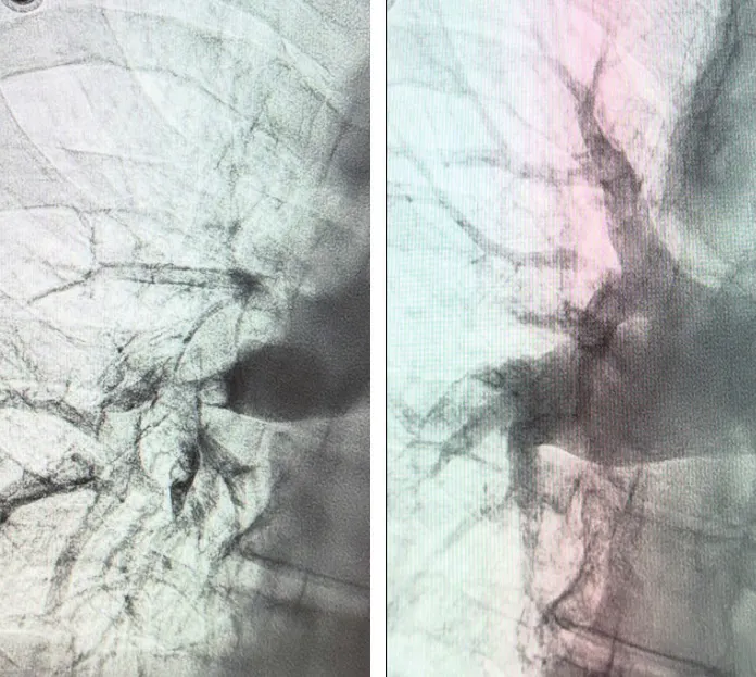
Figure 2. Pre- and post-thrombectomy pulmonary angiography.
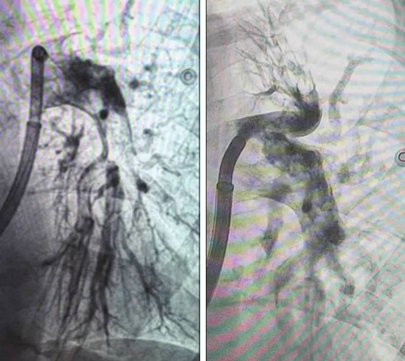
Figure 3. Pre- and post-thrombectomy pulmonary angiography.
Advertisement
Advertisement