Advertisement
Advertisement
2026 Digital Exclusive No. 2
Key Determinants of Escalation to MCS in Acute RV Failure
Experts share how hemodynamics, end-organ dysfunction, and underlying pathophysiology inform timely escalation to mechanical circulatory support in acute right ventricular failure.
With Ahmed Zayed, MBBCh; Spencer D. Liu, MD, FACC, FSCAI; and Marwan F. Jumean, MD, FACC, FSCAI


Right ventricular (RV) failure is a problem with no clean algorithm to follow; in many ways, it is a clinical art. In cardiogenic shock complicating acute myocardial infarction (AMI), RV failure carries mortality north of 60%, and once both ventricles are involved, 2-year survival drops to about 23% compared to 71% with isolated left-sided failure.1,2 Acute RV failure is often part of a bigger picture of a failing heart, while truly isolated RV failure from a massive pulmonary embolism (PE) or acute pulmonary hypertension is less common but can spiral just as fast.
Unlike many areas of practice where algorithms and randomized trials dictate clear decision points, RV failure demands a synthesis of hemodynamic data, imaging findings, end-organ markers, and occasionally clinical intuition. Delineating the primary etiology of RV failure fundamentally shapes both the trajectory and the therapeutic strategy. Normal RV function is governed by systemic venous return, pulmonary artery vascular resistance, and native contractility. What makes RV failure so tricky is that the right ventricle does not behave like the left ventricle. It handles volume reasonably well but does not tolerate acute pressure overload, and because its contractility depends on both the free wall and the interventricular septum, chronic left-sided disease often drags the RV down with it.3
Conceptually, the management of acute RV failure is similar to left ventricular (LV) failure and is one that we all know: preload, afterload, and contractility. Unfortunately, the choice of vasoactive support is not rooted in clinical evidence and is often anecdotal. As in all avenues of medicine, the choice of therapy should be individualized and determined by the available resources. Often, patients present with LV failure and are supported on a combination of medical and device therapy. We may think the left side is well supported and the wedge looks reasonable, but the patient is still not improving. That is often the moment RV failure presents itself and the conversation shifts toward RV support.
What makes us think we need more support? It starts with a hemodynamic assessment. The pulmonary artery catheter (PAC) is indispensable in the management of RV failure. The first parameter we look at for prognostication and decision-making is the central venous pressure (CVP). It is easy to obtain, does not necessitate the need for a PAC, and can be obtained from an indwelling central line. A CVP that stays elevated despite acceptable urine output and what should be adequate LV support is usually the first warning sign. The CVP-to-wedge ratio adds another layer: once that is above 0.9, we know the right ventricle is a key player. Pulmonary artery pulsatility index (PAPi) is another helpful tool, although you must be careful when severe pulmonary hypertension confounds the calculation. Finally, what tips the scale most often is not a single number but the metabolic picture. When the creatinine starts creeping up and liver function tests and bilirubin rise or the patient is not thriving, those are signs that end-organ perfusion is failing and a decision must be made regarding upgrading support. This is usually the driving force to escalate support even when the hemodynamics look borderline. Echocardiographic findings and risk predictor scores are useful, but more for risk stratification than for dictating the real-time decision to go mechanical.
Once we have made the decision to escalate, device selection comes down to the clinical scenario and, just as importantly, what your team can deploy confidently. RV support devices are not nearly as ubiquitous as LV devices, and operator comfort matters tremendously. If your institution runs extracorporeal membrane oxygenation (ECMO) well but is less experienced with dedicated RV assist devices (RVADs), you are better off with a well-managed ECMO circuit. If your facility does not have these capabilities at all, do not wait—transfer early. Before you add any device, have an exit strategy in mind. More devices mean more complications, and you must ask whether this patient has a realistic path to recovery, durable support, or advanced therapies.
Ultimately, the management of acute RV failure demands vigilance, physiologic precision, and decisiveness. Although prospective, randomized evidence comparing escalation strategies and device platforms remains an urgent need, the principles guiding our practice are consistent: Phenotype early, optimize aggressively, and escalate without hesitation when the clinical trajectory signals that medical therapy has reached its ceiling.
1. Harjola VP, Mebazaa A, Čelutkienė J, et al. Contemporary management of acute right ventricular failure: a statement from the Heart Failure Association and the Working Group on Pulmonary Circulation and Right Ventricular Function of the European Society of Cardiology. Eur J Heart Fail. 2016;18:226-241. doi: 10.1002/ejhf.478
2. Mehta SR, Eikelboom JW, Natarajan MK, et al. Impact of right ventricular involvement on mortality and morbidity in patients with inferior myocardial infarction. J Am Coll Cardiol. 2001;37:37-43. doi: 10.1016/S0735-1097(00)01089-5
3. Konstam MA, Kiernan MS, Bernstein D, et al. Evaluation and management of right-sided heart failure: a scientific statement from the American Heart Association. Circulation. 2018;137:e578-e622. doi: 10.1161/CIR.0000000000000560

Acute RV failure remains one of the most challenging syndromes in cardiovascular medicine due to its heterogeneous etiologies, variable presentation, and high mortality. Unlike LV failure, where robust evidence guides mechanical support strategies, the role of temporary mechanical circulatory support (MCS) in RV failure remains less well defined and often relies on careful integration of clinical judgment, hemodynamic assessment, and pathophysiology. The decision to escalate from medical therapy to mechanical RV support requires a nuanced understanding of the underlying cause, the severity of hemodynamic compromise, and the likelihood of reversibility.1-4
CHALLENGES IN DIAGNOSIS OF ACUTE RV FAILURE
A central difficulty in managing acute RV failure is establishing an early and accurate diagnosis. RV failure is not always clinically obvious and often presents in the context of complex systemic illness, such as AMI, septic shock, PE, or postcardiotomy states. Diagnosis requires alignment of four domains2,4-6:
- Clinical context: Patients may present with hypotension, elevated jugular venous pressure, hepatomegaly, peripheral edema, and signs of low cardiac output such as altered mentation or oliguria. However, these findings are often nonspecific and may overlap with left-sided or distributive shock.
- Biochemical markers: Elevated lactate levels indicate tissue hypoperfusion, while increases in liver enzymes (alanine transaminase/aspartate transaminase) and creatinine suggest end-organ congestion and impaired perfusion. Cardiac biomarkers such as troponin and B-type natriuretic peptide may indicate myocardial involvement but are not specific for RV failure.
- Imaging markers: Echocardiography is an essential diagnostic tool, but it may be technically limited by acoustic windows and the sonographer. Echocardiographic findings suggestive of RV failure include RV dilation (RV/LV ratio > 1), reduced fractional area change, decreased tricuspid annular plane systolic excursion, and septal flattening resulting in a “D-shaped” septum. However, these findings must be interpreted in the clinical context, particularly when distinguishing between acute and chronic RV pressure overload.5
- Invasive hemodynamics: Right-heart catheterization often provides critical diagnostic and physiologic insight. Hemodynamic findings suggestive of RV failure include elevated right atrial pressure (RAP), a low PAPi, reduced cardiac index, and a disproportionate elevation in RAP relative to pulmonary capillary wedge pressure. Additional findings may include a low pulmonary capillary wedge pressure and reduced RV stroke work and stroke work index.1,3,7
WHEN TO ESCALATE FROM MEDICAL THERAPY TO MECHANICAL SUPPORT
Initial management of acute RV failure focuses on optimizing preload, reducing afterload, and enhancing contractility. Strategies include volume management, vasopressors (eg, norepinephrine), inotropes (eg, dobutamine, milrinone), and pulmonary vasodilators (eg, inhaled nitric oxide). Escalation to mechanical support should be considered when these measures fail to achieve hemodynamic stability or when rapid deterioration is anticipated.
There are five key factors influencing the decision to escalate therapy2-4,8,9:
- Degree of hemodynamic collapse: Society for Cardiovascular Angiography & Interventions (SCAI) stage C or progression to a higher SCAI stage—characterized by persistent hypotension, rising lactate levels, worsening end-organ dysfunction (renal or hepatic), and inability to maintain adequate cardiac output despite escalating pharmacologic support—is a strong indicator for early escalation to MCS.
- Failure of medical therapy: Refractory shock despite optimized preload, high-dose vasopressors and inotropes, and pulmonary vasodilators suggests that the RV is unable to generate sufficient forward flow.2,8
- Evidence of end-organ injury: Rising bilirubin, transaminases, and creatinine indicate venous congestion and hypoperfusion. Early initiation of mechanical support may prevent irreversible injury.3,4
- Reversibility of underlying etiology: MCS is most beneficial when the underlying cause is reversible, such as acute RV infarction, myocarditis, or postcardiotomy stunning. In contrast, irreversible pathology, such as an interstitial lung disease flare, may limit its benefit unless used as a bridge to advanced therapies.2,4
- Timing—earlier is better: Delays in escalation often result in a downward spiral of multiorgan failure. Early deployment of mechanical support, before the onset of profound shock and organ dysfunction, may be associated with favorable outcomes; however, robust studies supporting this approach are lacking. A general rule of thumb is that if the vasoactive-inotropic score is > 5 or is rising, MCS support should be considered.3,4
PATHOPHYSIOLOGY-DRIVEN APPROACH TO MECHANICAL SUPPORT
A critical determinant of mechanical RV support effectiveness is the underlying mechanism of RV failure. Broadly, RV failure can be categorized into acute RV contractile failure and acute increases in RV afterload. Acute RV contractile failure may occur in acute RV myocardial infarction, myocarditis, and postcardiotomy RV dysfunction. In these scenarios, the primary issue is impaired RV contractility with relatively preserved pulmonary vascular resistance. Percutaneous RVADs are particularly effective in this setting, as they bypass the failing RV by directly pumping blood from the right atrium or vena cava into the pulmonary artery, thereby increasing pulmonary blood flow, improving LV preload, and enhancing systemic cardiac output.
Clinical experience suggests that patients with acute RV infarction derive the maximum benefit from percutaneous RVAD support, particularly when instituted early, before the development of irreversible ischemic damage and end-organ dysfunction.2,4
Acute increases in RV afterload, or pressure overload, occur in conditions such as massive or submassive PE, severe acute respiratory distress syndrome (ARDS), and acute pulmonary hypertension crises. In these settings, RV failure arises not from intrinsic contractile dysfunction but from a sudden increase in afterload. In these cases, an RVAD may not be effective because:4,6
- The device must pump against a markedly elevated pulmonary vascular resistance
- Forward flow remains limited despite mechanical assistance
- RV decompression does not address the primary issue of high afterload
In contrast, venoarterial (VA)-ECMO is often more appropriate in this scenario as it:2,4
- Bypasses both RV and pulmonary circulation
- Provides systemic perfusion independent of pulmonary vascular resistance
- Allows time to treat the underlying pathology, such as thrombolysis for PE or lung-protective ventilation for ARDS4,6
Thus, differentiating between contractile failure and afterload-driven failure is essential in selecting the appropriate support modality.
Available percutaneous RVAD systems, including dual-lumen cannula systems (eg, TandemHeart RVAD [LivaNova]) and pump-based platforms (eg, Impella RP and RP Flex [Abiomed]), provide effective short-term support but are not without limitations. Most percutaneous RVADs are designed for short-term use (days to 1-2 weeks) due to risks such as hemolysis, cannula-related complications, infection, thrombosis, and device migration or slippage from the pulmonary artery into the right ventricle. As a result, their use is generally limited to bridge-to-recovery, bridge-to-decision, or bridge-to-durable support strategies, including surgical RVAD placement. Complication rates increase with longer support duration and may include access-site bleeding, vascular injury, device malfunction, and arrhythmias. Therefore, careful patient selection and early planning for an exit strategy are essential when percutaneous RVAD support is considered.3
It is important to recognize that, unlike left-sided mechanical support, high-quality randomized data guiding RV support remain limited. Nonetheless, several key studies and registries have informed current clinical practice. The RECOVER RIGHT study evaluated percutaneous RVAD support using the Impella RP in patients with acute RV failure and demonstrated the feasibility of percutaneous RV support, rapid hemodynamic improvement, and an acceptable safety profile in selected patients. Although nonrandomized, this study provided clinical evidence supporting the use of percutaneous RVADs in acute settings.10
Despite insights from case reports and expert consensus, no formal guidelines define precise timing or patient selection for mechanical RV support, leaving clinicians heavily reliant on physiology-based decision-making. A practical approach to escalation is summarized in Figure 1.5,11
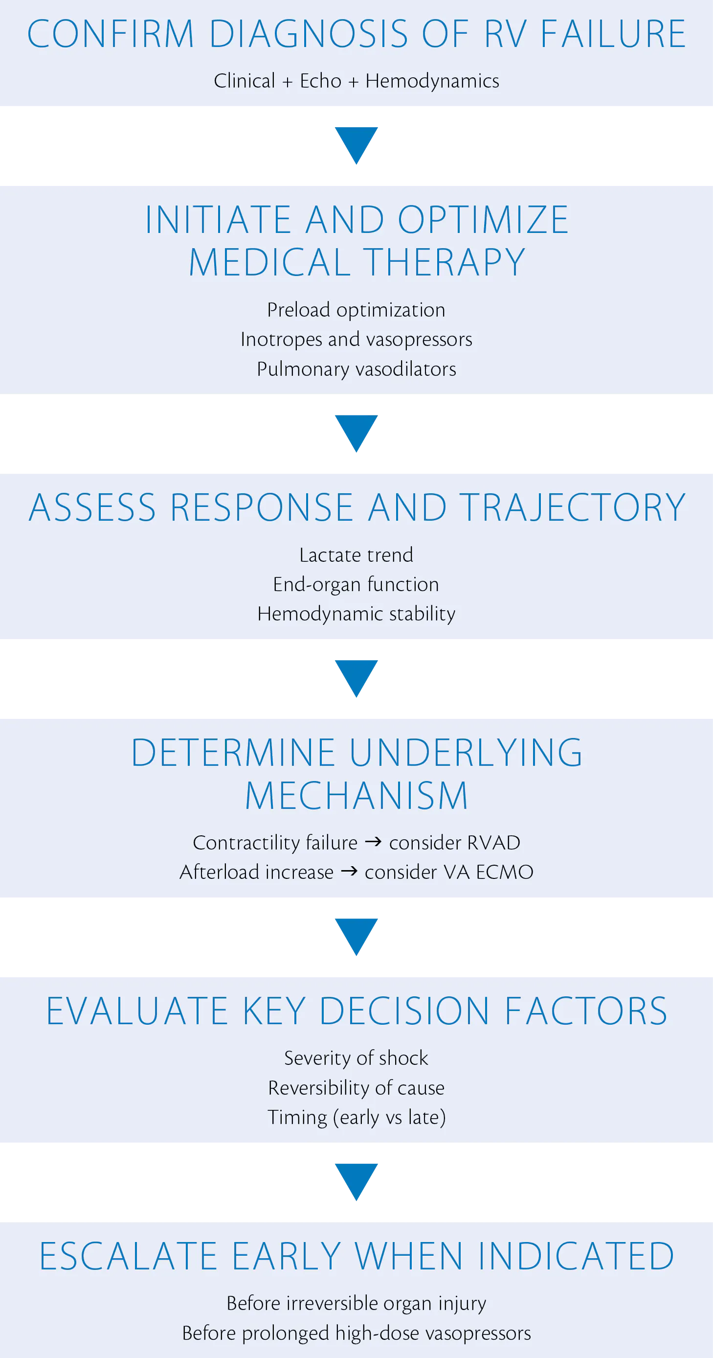
Figure 1. Clinical decision pathway for acute RV failure. RVAD, right ventricular assist device; VA-ECMO, venoarterial extracorporeal membrane oxygenation.2,4
CONCLUSION
The management of acute RV failure requires a careful balance between timely intervention and appropriate patient selection. Escalation from medical therapy to MCS should not be delayed until profound shock ensues, as outcomes worsen significantly once multiorgan failure develops. Instead, early recognition of deteriorating hemodynamics combined with a clear understanding of underlying pathophysiology should guide intervention.
Percutaneous RVAD support is most effective in states of acute RV contractile failure, such as RV infarction, where rapid restoration of forward flow and systemic perfusion can be achieved. In contrast, conditions characterized by elevated pulmonary vascular resistance often require more comprehensive support with VA-ECMO.2,4
Although high-quality evidence remains limited and formal guidelines are lacking, emerging data and growing clinical experience increasingly support a proactive, physiology-driven approach. Ultimately, early deployment of temporary MCS in the appropriate patient—before irreversible injury develops—remains the most important determinant of success in treating acute RV failure.5,8,11
1. Haddad F, Hunt SA, Rosenthal DN, Murphy DJ. Right ventricular function in cardiovascular disease, part I: anatomy, physiology, aging, and functional assessment of the right ventricle. Circulation. 2008;117:1436-1448. doi: 10.1161/CIRCULATIONAHA.107.653576
2. Houston BA, Brittain EL, Tedford RJ. Right ventricular failure. N Engl J Med. 2023;388:1111-1125. doi: 10.1056/NEJMra2207410
3. Vallabhajosyula S, Kumar M, Pandompatam G, et al. Prognostic impact of isolated right ventricular dysfunction in sepsis and septic shock: an 8-year historical cohort study. Ann Intensive Care. 2017;7:94. doi: 10.1186/s13613-017-0319-9
4. Giannakoulas G, Farmakis IT, Hobohm L, et al. Acute right ventricular failure: pathophysiology, aetiology, assessment, and management. Eur Heart J. 2025;46:2520-2535. doi: 10.1093/eurheartj/ehaf215
5. Rudski LG, Lai WW, Afilalo J, et al. Guidelines for the echocardiographic assessment of the right heart in adults: a report from the American Society of Echocardiography endorsed by the European Association of Echocardiography, a registered branch of the European Society of Cardiology, and the Canadian Society of Echocardiography. J Am Soc Echocardiogr. 2010;23:685-713. doi: 10.1016/j.echo.2010.05.010
6. Asakage A, Bækgaard J, Mebazza A, Deniau B. Management of acute right ventricular failure. 2023;20:218-229. doi: 10.1007/s11897-023-00601-5.
7. Damman K, van Deursen VM, Navis G, et al. Increased central venous pressure is associated with impaired renal function and mortality in a broad spectrum of patients with cardiovascular disease. J Am Coll Cardiol. 2008;53:582-588. doi: 10.1016/j.jacc.2008.08.080
8. Harjola VP, Mebazaa A, Čelutkienė J, et al. Contemporary management of acute right ventricular failure: a statement from the Heart Failure Association and the Working Group on Pulmonary Circulation and Right Ventricular Function of the European Society of Cardiology. Eur J Heart Fail. 2016;18:226-241. doi: 10.1002/ejhf.478
9. Bakar SN, Jia S, Smith SJ. Right ventricular failure management. Curr Opin Cardiol. 2019;34:213-217. doi: 10.1097/HCO.0000000000000595
10. Anderson MB, Goldstein J, Milano C, et al. Benefits of a novel percutaneous ventricular assist device for right heart failure: the RECOVER RIGHT study. J Heart Lung Transplant. 2015;34:1549-1560. doi:10.1016/j.healun.2015.08.018
11. Mehra MR, Park MH, Landzberg MJ, et al. Right heart failure: toward a common language. J Heart Lung Transplant. 2014;33:123-126. doi: 10.1016/j.healun.2013.10.015
Advertisement
Advertisement
Latest Digital Exclusives