
Advertisement
Advertisement
2026 Digital Exclusive No. 1
Coronary Bifurcation Imaging:
What I’m Looking for and How I Do It
The technical advantages of a systematic strategy for intravascular imaging for bifurcation PCI and how and why to use it before stenting, after stenting, and for left main bifurcation.
Bifurcation percutaneous coronary intervention (PCI) can be technically challenging and represents up to 20% of interventions performed in contemporary clinical practice.1,2 Bifurcation PCI is also associated with higher rates of cardiovascular events and a lower probability of procedural success as compared with nonbifurcation PCI. Fortunately, as a result of numerous clinical trials over the past 2 decades, efforts have been made to optimize the strategy for these complex interventions.3-5
COMING OF AGE: INTRAVASCULAR ULTRASOUND
Utilization of intravascular ultrasound (IVUS) did not significantly increase until the second decade of the 21st century, in response to data demonstrating improved PCI outcomes with its use.6 The two most significant randomized trials are ULTIMATE and RENOVATE-COMPLEX-PCI. The former showed lower rates of target vessel failure (TVF) and thrombosis in all-comers receiving PCI with the use of IVUS compared with angiographic guidance.7 This result was sustained at 3 years.8 In RENOVATE-COMPLEX-PCI, there was a lower rate of TVF after PCI of complex lesions with intravascular imaging (IVI) compared with angiography.9 Although the use of either IVUS or optical coherence tomography (OCT) was allowed in the imaging arm, the majority (73%) of operators used IVUS. Accordingly, the American College of Cardiology/American Heart Association guidelines for coronary revascularization and for management of patients with acute coronary syndrome recommend IVUS for PCI in the left main coronary artery (LMCA) or for complex lesions, including bifurcation.10,11
A RACE BETWEEN LIGHT AND SOUND: OCT VERSUS IVUS
Prior to more recent trials, several randomized trials did show noninferiority of OCT to IVUS.12,13 However, only IVUS outperformed angiography head-to-head in a randomized trial, whereas OCT had similar rates of TVF compared to angiography.7,14 Nonetheless, two recent trials, RENOVATE-COMPLEX-PCI and OCTOBER, have re-established OCT as a contender in the IVI arena,9,15 and more recent guidelines do not prescribe one over the other.10 Indeed, the recent OCTOBER trial demonstrated lower TVF with OCT in true bifurcation lesions.15
WHY USE IVI FOR BIFURCATION PCI?
The technical advantages of IVI over angiography alone in bifurcation PCI are many. IVUS allows for the following:
- Ensuring adequate lesion preparation prior to stent deployment and understanding of lesion morphology
- Precise evaluation of side branch disease and risk of side branch compromise, including understanding the minimal lumen area of the side branch and potential need for two-stent approach
- Precise stent sizing (especially relevant for stents crossing from proximal to distal main branch)
- Ensuring adequate stent expansion and apposition
PRE-STENT IVI
Prior to Lesion Preparation
The primary benefits of performing IVI prior to any lesion preparation are, arguably, (1) the ability to understand lesion morphology, including calcification and the mechanism of in-stent restenosis (ISR); and (2) for side branch evaluation to assist in the decision between provisional or upfront two-stent strategies. Both IVUS and OCT can be effective for identifying severe calcification, defined as > 270° arc of calcium extending at least 5 mm down the vessel or a full 360° of calcium. The presence of severe calcification may alter the PCI plan in favor of early atherectomy given the limited efficacy of balloon angioplasty in treating severely calcified lesions.16 Figure 1 shows examples of calcification on IVUS.
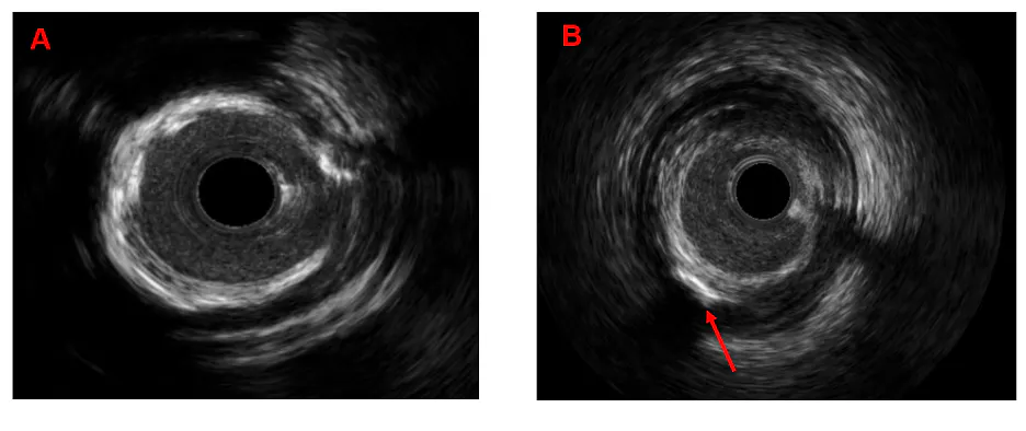
Figure 1. A case of severe superficial calcium extending almost 360° inside the vessel lumen (A). Mild calcification deep to the intima with characteristic shadowing behind the calcium (B).
Additionally, understanding the mechanism of ISR may also dictate the PCI strategy. For example, neointimal hyperplasia is best addressed with drug-eluting stents and/or angioplasty, whereas underexpansion is best treated with re-expansion of the stent.17 Guidelines recommend IVI to aid in evaluating stent failure (Figure 2).11
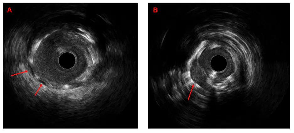
Figure 2. A case of stent failure secondary to malapposition (image obtained after initial balloon angioplasty) (A). A case of severe ISR secondary to neointimal hyperplasia (B).
Finally, IVI can be used to evaluate the side branch prior to disrupting it, especially in cases where there is uncertainty regarding the risk of side branch compromise and need for a planned two-stent strategy (ie, whether there is ostial side branch disease). IVI can confirm the side branch diameter (for the purposes of understanding its significance) and evaluate the DEFINITION criteria for complex bifurcation disease, including the presence of > 90% ostial disease in the side branch or 10-mm disease length. Further, IVUS allows precise measurement of the side branch angle.18,19 In cases of complex bifurcation disease, an upfront two-stent strategy may be preferred.19 The so-called “eyebrow” sign on longitudinal reconstruction—when the main branch and side branch are initially parallel before the latter changes course—is also predictive of side branch compromise.20 However, it is important to note that in heavily calcified lesions, performing IVI before lesion preparation may not be possible or can lead to complications or entrapment of the IVUS or OCT catheters. It is important to never push IVI catheters against resistance in lesions prior to preparation.
After Lesion Preparation
Postlesion preparation IVI can be useful for confirming adequate lesion preparation, identifying a landing zone, and sizing the diameter and length of the landing zone for precise stent selection.
For heavily calcified bifurcation lesions where lesion calcium modification was necessary, it may be particularly beneficial to perform IVI of both the main and side branch. This ensures adequate lesion preparation prior to pursuing a two-stent or provisional strategy (Figure 3).
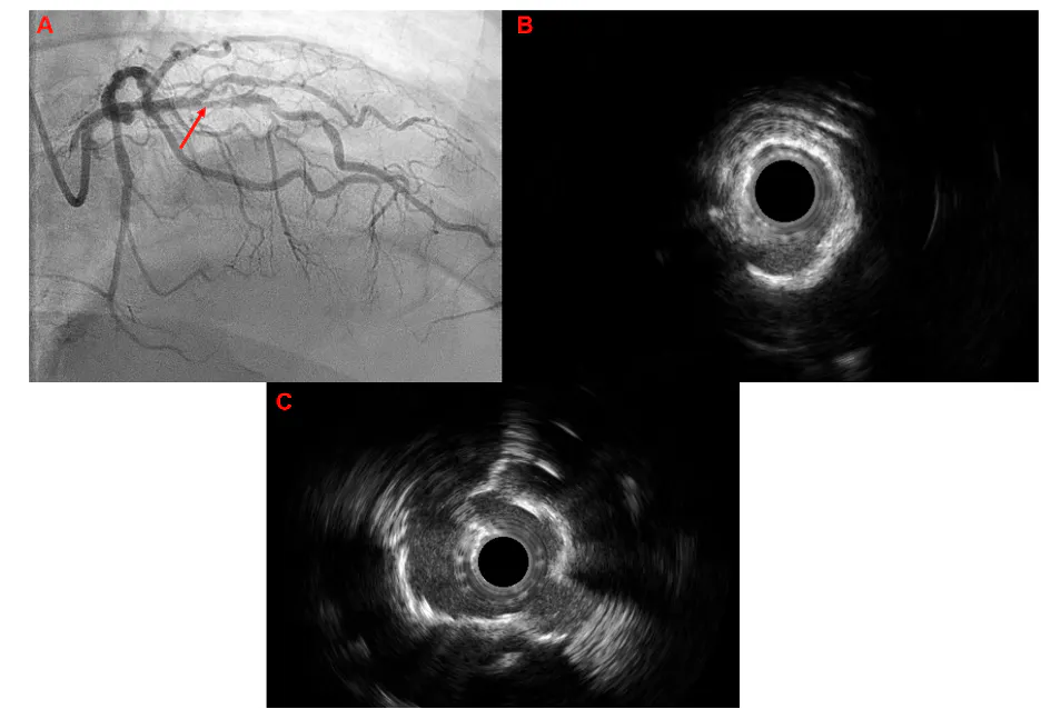
Figure 3. A highly calcified mid LAD lesion (A). IVUS showing 360° calcification at the lesion (B). Fracture of the calcium after rotational atherectomy (C).
Arguably, the most critical application of IVI is to ensure adequate stent expansion. Pre-stent imaging allows for appropriate sizing of the stent, especially in cases of bifurcation PCI where there is often a significant size mismatch between the distal and proximal landing zones in the main branch (Figure 4). This ensures selection of a stent large enough to perform the proximal optimization technique but small enough for the distal vessel.
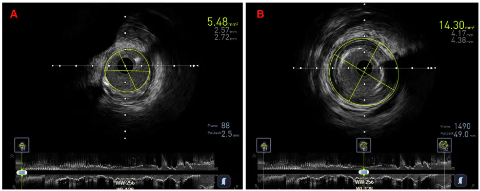
Figure 4. Sizing of the distal vessel reference vessel (A) and sizing of the proximal reference vessel (B), significantly different in size and necessitating two overlapping stents.
POST-STENT IVI
IVI after stenting has the advantage of confirming adequate stent expansion and apposition, excluding significant edge dissection or plaque shift, and confirming adequate lesion coverage, including evaluating for geographic miss.
Notably, randomized trials of IVI in PCI all mandated post-stent imaging to confirm optimal stent deployment.7,9 At the least, post-stent imaging is useful to confirm optimal stent deployment in the main branch and selectively in the side branch in cases where there is a poor angiographic result with stent underexpansion, concerns regarding stent length and adequate coverage of the lesion, or angiographic evidence of edge disease, including significant edge dissection.
LEFT MAIN BIFURCATION
Challenges to interventions in the LMCA include a high incidence of calcification and bifurcation lesions that may result in hemodynamic or myocardial compromise of myocardial territories supplied by the left anterior descending (LAD) or left circumflex (LCx) arteries (Figure 5).
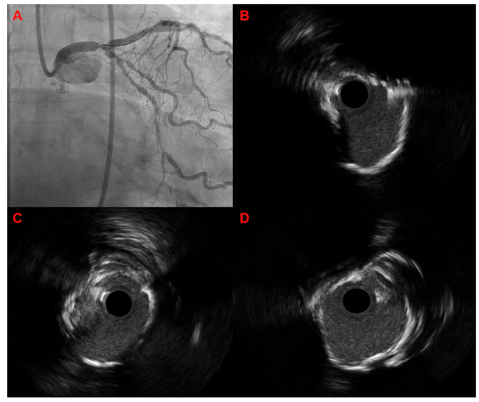
Figure 5. A severely stenotic LMCA (A) associated with significant calcification on IVUS (B). Significant, calcific disease at the ostium of the LAD (C) and LCx (D) arteries.
There are four contemporary trials that have evaluated PCI outcomes in the LMCA, with the most notable being EXCEL and NOBLE.21,22
IVI should be used in all left main or left main bifurcation lesions given the risk of inadequate lesion preparation or stent malapposition compromising significant myocardial territory. Guidelines recommend IVI to support LMCA PCI, both for evaluating indeterminate lesions and for procedural guidance.10,11 IVUS area measurements in the LMCA can be used to determine stenosis severity (a minimum lumen area < 6-7.5 mm2 is generally considered significant), and these measurements correlate well with coronary physiology.11
CONCLUSION
Bifurcation lesions present challenging scenarios for PCI. Nonetheless, careful and systematic utilization of IVI can eliminate some of the uncertainty with bifurcation PCI and improve cardiovascular outcomes.
1. Riley RF, Henry TD, Mahmud E, et al. SCAI position statement on optimal percutaneous coronary interventional therapy for complex coronary artery disease. Catheter Cardiovasc Interv. 2020;96:346-362. doi: 10.1002/ccd.28994
2. Latib A, Colombo A. Bifurcation disease: what do we know, what should we do? JACC Cardiovasc Interv. 2008;1:218-226. doi: 10.1016/j.jcin.2007.12.008
3. Lassen JF, Holm NR, Banning A, et al. Percutaneous coronary intervention for coronary bifurcation disease: 11th consensus document from the European Bifurcation Club. EuroIntervention. 2016;12:38-46. doi: 10.4244/EIJV12I1A7
4. Steigen TK, Maeng M, Wiseth R, et al; Nordic PCI Study Group. Randomized study on simple versus complex stenting of coronary artery bifurcation lesions: the Nordic bifurcation study. Circulation. 2006;114:1955-1961. doi: 10.1161/CIRCULATIONAHA.106.664920
5. Chen SL, Zhang JJ, Han Y, et al. Double kissing crush versus provisional stenting for left main distal bifurcation lesions: DKCRUSH-V randomized trial. J Am Coll Cardiol. 2017;70:2605-2617. doi: 10.1016/j.jacc.2017.09.1066
6. Stein EJ, Mesenbring E, Smith T, et al. Intravascular imaging as a performance measure for percutaneous coronary intervention. Circ Cardiovasc Interv. 2025;18:e014528. doi: 10.1161/CIRCINTERVENTIONS.124.014528.
7. Zhang J, Gao X, Kan J, et al. Intravascular ultrasound versus angiography-guided drug-eluting stent implantation: the ULTIMATE trial. J Am Coll Cardiol. 2018;72:3126-3137. doi: 10.1016/j.jacc.2018.09.013
8. Gao XF, Ge Z, Kong XQ, et al; ULTIMATE Investigators. 3-Year outcomes of the ULTIMATE trial comparing intravascular ultrasound versus angiography-guided drug-eluting stent implantation. JACC Cardiovasc Interv. 2021;14:247-257. doi: 10.1016/j.jcin.2020.10.001
9. Lee JM, Choi KH, Song YB, et al; RENOVATE-COMPLEX-PCI Investigators. Intravascular imaging-guided or angiography-guided complex PCI. N Engl J Med. 2023;388:1668-1679. doi: 10.1056/NEJMoa2216607
10. Rao SV, O'Donoghue ML, Ruel M, et al. 2025 ACC/AHA/ACEP/NAEMSP/SCAI guideline for the management of patients with acute coronary syndromes: a report of the American College of Cardiology/American Heart Association joint committee on clinical practice guidelines. Circulation. 2025;151:e771-e862. Published corrections appear in Circulation. 2025;151:e865 and Circulation. 2025;151:e1098. doi: 10.1161/CIR.0000000000001309
11. Writing Committee Members; Lawton JS, Tamis-Holland JE, Bangalore S, et al. 2021 ACC/AHA/SCAI guideline for coronary artery revascularization: a report of the American College of Cardiology/American Heart Association joint committee on clinical practice guidelines. J Am Coll Cardiol. 2022;79:e21-e129. Published correction appears in J Am Coll Cardiol. 2022;79:1547 and J Am Coll Cardiol. 2024;84:771. doi: 10.1016/j.jacc.2021.09.006
12. Ali ZA, Karimi Galougahi K, Maehara A, et al. Outcomes of optical coherence tomography compared with intravascular ultrasound and with angiography to guide coronary stent implantation: one-year results from the ILUMIEN III: OPTIMIZE PCI trial. EuroIntervention. 2021;16:1085-1091. doi: 10.4244/EIJ-D-20-00498
13. Kubo T, Shinke T, Okamura T, et al; OPINION Investigators. Optical frequency domain imaging vs. intravascular ultrasound in percutaneous coronary intervention (OPINION trial): one-year angiographic and clinical results. Eur Heart J. 2017;38:3139-3147. doi: 10.1093/eurheartj/ehx351
14. Ali ZA, Landmesser U, Maehara A, et al; ILUMIEN IV Investigators. Optical coherence tomography-guided versus angiography-guided PCI. N Engl J Med. 2023;389:1466-1476. doi: 10.1056/NEJMoa2305861
15. Holm NR, Andreasen LN, Neghabat O, et al; OCTOBER Trial Group. OCT or angiography guidance for PCI in complex bifurcation lesions. N Engl J Med. 2023;389:1477-1487. doi: 10.1056/NEJMoa2307770
16. Riley RF, Patel MP, Abbott JD, et al. SCAI expert consensus statement on the management of calcified coronary lesions. J Soc Cardiovasc Angiogr Interv. 2024;3:101259. doi: 10.1016/j.jscai.2023.101259
17. Klein LW, Nathan S, Maehara A, et al. SCAI expert consensus statement on management of in-stent restenosis and stent thrombosis. J Soc Cardiovasc Angiogr Interv. 2023;2:100971. doi: 10.1016/j.jscai.2023.100971
18. Ryu HM, Kim BK, Kim JS, et al. Comparison between measured and calculated length of side branch ostium in coronary bifurcation lesions with intravascular ultrasound. Yonsei Med J. 2012;53:680-684. doi: 10.3349/ymj.2012.53.4.680
19. Chen SL, Sheiban I, Xu B, et al. Impact of the complexity of bifurcation lesions treated with drug-eluting stents: the DEFINITION study (definitions and impact of complex bifurcation lesions on clinical outcomes after percutaneous coronary intervention using drug-eluting stents). JACC Cardiovasc Interv. 2014;7:1266-1276. doi: 10.1016/j.jcin.2014.04.026
20. Suárez de Lezo J, Medina A, Martín P, et al. Predictors of ostial side branch damage during provisional stenting of coronary bifurcation lesions not involving the side branch origin: an ultrasonographic study. EuroIntervention. 2012;7:1147-1154. doi: 10.4244/EIJV7I10A185
21. Stone GW, Sabik JF, Serruys PW, et al; EXCEL Trial Investigators. Everolimus-eluting stents or bypass surgery for left main coronary artery disease. N Engl J Med. 2016;375:2223-2235. Published correction appears in N Engl J Med. 2019;381:1789. doi: 10.1056/NEJMoa1610227
22. Mäkikallio T, Holm NR, Lindsay M, et al; NOBLE study investigators. Percutaneous coronary angioplasty versus coronary artery bypass grafting in treatment of unprotected left main stenosis (NOBLE): a prospective, randomised, open-label, non-inferiority trial. Lancet. 2016;388:2743-2752. Published correction appears in Lancet. 2016;388:2742. doi: 10.1016/S0140-6736(16)32052-9
Advertisement
Advertisement

Latest Digital Exclusives